DRK7020 Particle Image Analyzer
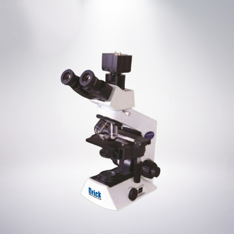
The drk-7020 particle image analyzer combines traditional microscopic measurement methods with modern image technology. It is a particle analysis system that uses image methods for particle morphology analysis and particle size measurement. It consists of an optical microscope, digital CCD camera and particle Image processing and analysis software composition. The system uses a dedicated digital camera to shoot the particle images of the microscope and transmit them to the computer. The image is processed and analyzed through a dedicated particle image processing and analysis software. It has the characteristics of intuitiveness, vividness, accuracy and wide test range. The morphology of the particles can be observed, and the analysis results such as the particle size distribution can also be obtained.
Technical Parameter
Measuring range: 1~3000 microns
Maximum optical magnification: 1600 times
Maximum resolution: 0.1 micron/pixel
Accuracy error: <±3% (national standard material)
Repeatability deviation: <±3% (national standard material)
Data output: perimeter distribution, area distribution, long diameter distribution, short diameter distribution, circumference equivalent diameter distribution, area equivalent diameter distribution, Feret diameter distribution, length to short diameter ratio, middle (D50), effective particle size (D10), limit Particle size (D60, D30, D97), number length average diameter, number area average diameter, number volume average diameter, length area average diameter, length volume average diameter, area volume average diameter, uneven coefficient, curvature coefficient.
Configuration parameters (configuration 1 domestic microscope) (configuration 2 imported microscope)
Trinocular Biological Microscope: Plan Eyepiece: 10×, 16×
Achromatic objective lens: 4×, 10×, 40×, 100× (oil)
Total magnification: 40×-1600×
Camera: 3 million pixel digital CCD (standard C-mount lens)
Scope of application
It is suitable for particle size measurement, morphology observation and analysis of various powder particles such as abrasives, coatings, non-metallic minerals, chemical reagents, dust, and fillers.
Software function and report output format
1. You can perform multiple processing on the image: such as: image enhancement, image superimposition, partial extraction, directional amplification, contrast, brightness adjustment and other dozens of functions.
2. It has basic measurement of dozens of geometric parameters such as roundness, curve, perimeter, area, and diameter.
3. The distribution diagram can be drawn directly by linear or non-linear statistical methods according to multiple types of parameters such as particle size, size, area, shape, etc.
Products categories
-

Phone
-

E-mail
-

Whatsapp
-

Top


